

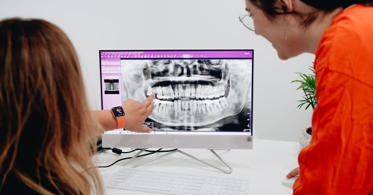
Extraction Complexity Assessment & Surgical Planning
Assess extraction complexity and generate customized surgical protocols for difficult teeth
What You Can Do
You can input radiographic findings, clinical presentation, and anatomical data to receive a comprehensive complexity assessment that identifies risk factors and generates customized surgical protocols. The skill integrates established classification systems (Pell & Gregory, Winter's classification) with proximity analysis to vital structures like the inferior alveolar nerve and maxillary sinus, producing evidence-based recommendations that reduce operative time, complications, and post-operative morbidity while protecting against medicolegal exposure.
Features
automatically classifies impacted molars and determines baseline surgical complexity
assesses proximity to inferior alveolar nerve, maxillary sinus, lingual plate, and other critical structures
evaluates calcification, fusion, curvature, and alveolar bone density to predict extraction resistance
categorizes cases into low, moderate, high, and extreme complexity tiers with corresponding surgical modifications
produces step-by-step surgical approaches, instrumentation recommendations, and complication prevention strategies
incorporates patient age, medical comorbidities, medications, and healing factors into treatment planning
generates evidence-based documentation suitable for informed consent and medicolegal protection
structures systematic evaluation for resident education and case-based surgical decision-making
Example Output
Example 1: Impacted Third Molar Assessment
Complexity Level: High (Pell III, Winter C, with IAN canal contact)
Key Risk Factors Identified:
- ✓ Deep impaction requiring significant bone removal
- ✓ Distal root angulation with curved root anatomy
- ✓ Intimate contact with IAN canal on panoramic and CBCT imaging
- ✓ Dense cortical bone requiring rotary instrumentation
Recommended Surgical Protocol:
- 2-3mm bone removal for inferior border clearance
- Sagittal split or high lingual extension retraction
- Piezoelectric bone reduction to protect nerve proximity
- Tooth sectioning (mesiodistal) to reduce extraction force
- Postoperative nerve assessment at 24 hours, 1 week, 3 months
Example 2: Retained Root Fragment with Sinus Proximity
Complexity Level: Moderate-High (3mm separation from sinus floor)
Key Risk Factors Identified:
- ✓ Separated tooth fragment with unclear remaining root length
- ✓ Maxillary sinus floor proximity requiring careful elevation technique
- ✓ Dense bone limiting instrumentation access
- ✓ Perforated sinus membrane risk during removal
Recommended Surgical Protocol:
- Limited flap elevation with sinus membrane-protective technique
- Careful bone removal under direct visualization
- Gentle elevation without rotational forces
- Consider CBCT imaging if fragment location unclear
- Post-extraction inspection for retained root particles
What's Included
- SKILL.md instruction file with complete extraction assessment framework:
- Radiographic Assessment Checklist: systematic review template for panoramic, periapical, and CBCT findings
- Complexity Classification Matrix: Pell & Gregory, Winter's, and proximity-based scoring system
- Risk Stratification Rubric: low/moderate/high/extreme tier definitions with surgical modifications
- Customizable Surgical Protocol Template: step-by-step approach selection based on complexity tier
- Systemic Risk Factor Worksheet: integration of patient medical history, medications, and healing considerations
- Informed Consent Documentation Guide: evidence-based language for pre-operative discussion and documentation
Who It's For
- Oral and maxillofacial surgeons — for pre-operative case assessment and surgical planning across all extraction complexity levels
- Oral surgery residents and fellows — for structured case evaluation, decision-making mentorship, and systematic surgical reasoning
- General dentists performing extractions — for risk assessment and referral decision-making on complex cases
- Dental specialists (periodontists, prosthodontists, orthodontists) — for pre-treatment extraction planning and complexity communication
- Surgical team leads — for consistent case assessment protocols and complication risk reduction across practice settings
Best For
- Impacted third molar assessment and treatment planning (all Pell & Gregory classifications)
- Difficult extractions involving retained roots, calcified teeth, or fused tooth anatomy
- Cases with proximal anatomical risk (inferior alveolar nerve canal, maxillary sinus, lingual plate proximity)
- Risk stratification before elective or urgent extractions requiring informed consent and medicolegal documentation
- Surgical protocol customization based on individual patient anatomy, pathology, and surgeon capability
- Resident and fellow education in systematic case evaluation and surgical decision-making frameworks
You might also like
You can systematically develop phase-by-phase orthodontic treatment protocols that synthesize diagnostic data, patient anatomy, and mechanical principles. This skill generates detailed appliance selection recommendations, extraction vs. non-extraction analyses, wire progression sequences, and clinical milestones to guide efficient execution from initial placement through retention for complex malocclusions requiring three-dimensional control.
 by comeonoliver
You can input detailed case information—including patient history, radiographic findings, clinical examination results, and anatomical considerations—and receive a comprehensive treatment analysis. The skill generates structured case summaries, identifies critical diagnostic elements, evaluates treatment options (conventional endodontic, surgical, or extraction), and produces documentation-ready prognosis assessments that support clinical decision-making and patient communication.
by comeonoliver
Stop bleeding money on forgotten subscriptions and negotiable expenses. This skill systematically dissects your spending, exposes hidden patterns you've overlooked, and generates a prioritized action plan that typically uncovers $2,000-$8,000+ in annual savings. You'll get specific negotiation templates, insider tips for lowering bills, and a complete expense audit that tells you exactly where your money is really going.
by comeonoliver
You can generate complete treatment plans for traditional fixed appliance cases by integrating diagnostic data, skeletal/dental analysis, and biomechanical principles. Claude helps you structure extraction vs. non-extraction decisions, design anchorage control strategies, sequence mechanics systematically, and create patient-facing narratives that explain your clinical rationale. This skill is designed for moderate-to-complex cases where defensible clinical reasoning and treatment efficiency optimization are critical.
by comeonoliver
This skill provides endodontists with a systematic protocol for assessing acute dental trauma cases, establishing urgency levels based on injury type and pulp vitality indicators, and creating documented baseline diagnostics that inform treatment decisions. You'll apply standardized assessment criteria across crown fractures, root fractures, luxation injuries, and avulsions—determining which cases need immediate intervention, which benefit from staged treatment, and which require specialist consultation or referral.
by comeonoliver
You can comprehensively analyze failed root canal cases to pinpoint why initial treatment didn't succeed—whether due to technical errors, anatomical challenges, or biological factors. Claude helps you develop systematic retreatment protocols, document clinical decision-making for peer review and medicolegal protection, and create patient-facing explanations of treatment failure mechanisms.
by comeonoliver
You can systematically assess acute dentoalveolar injuries using structured frameworks that classify fracture types, luxation stages, and avulsion status. The skill guides you through rapid determination of urgency tiers, identifies associated injuries requiring coordination with other specialties, and establishes evidence-based treatment timelines—all while generating standardized documentation for medico-legal purposes.
by comeonoliver
You can systematically evaluate patients with severe skeletal discrepancies and develop coordinated surgical treatment plans that integrate cephalometric analysis, surgical movement simulation, and patient communication strategies. This skill structures the entire pre-operative planning process—from baseline skeletal assessment through surgical consultation readiness—ensuring precise documentation and clear coordination with maxillofacial surgery partners.
by comeonoliver